Its Not All Bad if its Not All Around. Unmasking the Truth Beyond Myth.
Authors: Vibha Hayagreev, MD; Hardik Fichadiya, MD1; A. Chapa-Rodriguez, MD; Usman Younus, MD2
1 - Department of Cardiovascular Diseases, Cape Fear Valley Medical Center, Fayetteville, NC
2 - Department of Pulmonary and Critical Care, Cape Fear Valley Medical Center, Fayetteville, NC
Case
A 69-year-old male with a past medical history of atrial fibrillation/flutter and ischemic cardiomyopathy was admitted to the intensive care unit with a non-ST elevation myocardial infarction (NSTEMI), complicated by cardiogenic shock. A coronary angiogram revealed multi-vessel disease. The patient underwent coronary artery bypass grafting (CABG) along with mechanical circulatory support via a percutaneous micro axial pump (Impella 5.5). Post-operatively, the patient experienced episodes of ventricular tachycardia, which were managed with amiodarone and mexiletine. Despite treatment, he became hemodynamically unstable, with episodes of diastolic suction alarms and loss of flow from the Impella, leading to a rapid escalation of vasopressor support. Pulmonary artery catheterization showed the following hemodynamic parameters:
- Central venous pressure (CVP): 22 mmHg
- Pulmonary capillary wedge pressure (PCWP): 27 mmHg
- Cardiac index (CI): 1.78 L/min/m²
- SvO2: 50%
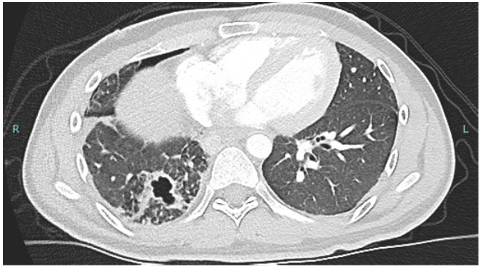
Transthoracic echocardiography (TTE) was difficult to perform due to the patient's post-operative condition but revealed that the Impella was abutting the septal wall. Despite repositioning, there was only a minimal improvement in flow. The patient's hemodynamic status continued to deteriorate. Given these findings, a transesophageal echocardiogram (TEE) was performed and showed:
Question
Based on the above image, what is the most likely diagnosis and the next best step in managing this patient ?
- Improper Impella position and repositioning of Impella
- Hypovolemic shock and fluid resuscitation
- Right ventricular (RV) failure and rescue with RV support
- Localized cardiac tamponade and mediastinal re-exploration with evacuation of hematoma
D. Localized cardiac tamponade and mediastinal re-exploration with evacuation of hematoma
Discussion
The diagnosis in this case is localized pericardial tamponade, and the next best step in management is mediastinal re-exploration with evacuation of the hematoma. Post-operative complications in cardiac surgery are varied and can include graft failure, bleeding, unstable arrhythmias, sepsis, and pulmonary embolism. Patients receiving systemic anticoagulation are at higher risk for pericardial hematoma, a potentially life-threatening complication.
Classic signs of tamponade, such as Beck’s triad (muffled heart sounds, pulsus paradoxus, and jugular venous distention), may not always be clinically apparent. Additionally, tamponade can be difficult to diagnose, particularly when the effusion is localized to the posterior or lateral aspects of the pericardium. This makes it poorly visualized on a TTE, especially in the post-operative period. Conventional tests, such as electrocardiogram and chest X-ray, may also be suboptimal in diagnosing this condition.
A large circumferential pericardial effusion or an acute smaller effusion may raise concerns for tamponade physiology, but it is important to remember that localized pericardial effusions can also lead to tamponade by causing regional chamber compression. This type of effusion, along with hematomas, thrombus, or masses, can result in cardiac tamponade.
TEE is a critical diagnostic tool in this setting, as it offers better image quality, especially in post-operative patients. It also facilitates improved visualization of the posterior heart, which is often challenging to assess with TTE. Given the patient's high risk of tamponade, early use of TEE can assist in rapid diagnosis, leading to timely interventions that can be lifesaving.
Conclusion
In patients with a high index of suspicion for cardiac tamponade, TEE should be performed promptly to confirm the diagnosis. Early intervention with mediastinal re-exploration and evacuation of hematoma is often required in these cases. Prompt decision-making and intervention are essential in preventing further hemodynamic deterioration and improving patient outcomes.
References
-
Nguyen HS, Nguyen HD, Vu TD. Pericardial effusion following cardiac surgery: A single-center experience. Asian Cardiovasc Thorac Ann. 2018;26(1):5–10.
-
Kuvin JT, Harati NA, Pandian NG, Bojar RM, Khabbaz KR. Postoperative cardiac tamponade in the modern surgical era. Ann Thorac Surg. 2002;74(4):1148–1153.
-
Ashikhmina EA, Schaff HV, Sinak LJ, Li Z, Dearani JA, Suri RM, Park SJ, Orszulak TA, Sundt TM. Pericardial effusion after cardiac surgery: Risk factors, patient profiles, and contemporary management. Ann Thorac Surg. 2010;89(1):112–118.
-
Pepi M, Muratori M, Barbier P, Doria E, Arena V, Berti M, Celeste F, Guazzi M, Tamborini G. Pericardial effusion after cardiac surgery: Incidence, site, size, and haemodynamic consequences. Br Heart J. 1994;72(4):327–331.
-
Ionescu A, Wilde P, Karsch KR. Localized pericardial tamponade: Difficult echocardiographic diagnosis of a rare complication after cardiac surgery. J Am Soc Echocardiogr. 2001;14(12):1220–1223.
-
Russo AM, O’Connor WH, Waxman HL. Atypical presentations and echocardiographic findings in patients with cardiac tamponade occurring early and late after cardiac surgery. Chest. 1993;104(1):71–77.