Reviewed By Allergy, Immunology & Inflammation Assembly
Submitted by
Lokesh Venkateshaiah, MD
Fellow
Division of Pulmonary, Critical Care and Sleep Medicine
Case Western Reserve University
Cleveland, Ohio
J. Daryl Thornton, MD MPH
Assistant Professor
Division of Pulmonary, Critical Care and Sleep Medicine
Case Western Reserve University
Cleveland, Ohio
Submit your comments to the author(s).
History
Physical Exam
Lab
Figures

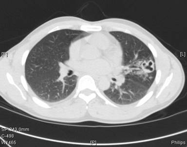
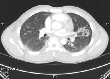
Fig 2: Chest computed tomography at presentation (4 months following initiation of antituberculous therapy)
References
- Meehan AM, Virk A, Swanson K, et al: Severe pleuropulmonary paragonimiasis 8 years after emigration from a region of endemicity. Clin Infect Dis 2002; 35:87-90.
- Yee B, Hsu J-I, Favour CB, et al: Pulmonary paragonimiasis in Southeast Asians living in the Central San Joaquin Valley. West J Med 1992; 156:423-425
- Procop, GW, Marty, AM, Scheck, DN, et al. North American paragonimiasis. A case report. Acta Cytol 2000; 44:75.
- Mariano, EG, Borja, SR, Vruno, MJ. A human infection with Paragonimus kellicotti (lung fluke) in the United States. Am J Clin Pathol 1986; 86:685.
- DeFrain, M, Hooker, R. North American paragonimiasis: case report of a severe clinical infection. Chest 2002; 121:1368.
- Castilla, EA, Jessen, R, Sheck, DN, Procop, GW. Cavitary mass lesion and recurrent pneumothoraces due to Paragonimus kellicotti infection: North American paragonimiasis. Am J Surg Pathol 2003; 27:1157.
- www.dpd.cdc.gov
- Mackie,T. Parasitic infections of the Lung; Chest 1948; 14; 894-905
- Heath, Harley W & Susan G Marshall. "Pleural Paragonimiasis In A Laotian Child. ." Pediatric Infectious Disease Journal 16(12)(1997): :1182-1185
- Im, JG, Whang, HY, Kim, WS, et al. Pleuropulmonary paragonimiasis: radiologic findings in 71 patients. AJR Am J Roentgenol 1992; 159:39.
- Singcharoen, T, Silprasert, W. CT findings in pulmonary paragonimiasis. J Comput Assist Tomogr 1987; 11:1101.
- Kim, TS, Han, J, Shim, SS, et al. Pleuropulmonary paragonimiasis: CT findings in 31 patients. AJR Am J Roentgenol 2005; 185:616.
- Tomita, M, Matsuzaki, Y, Nawa, Y, Onitsuka, T. Pulmonary paragonimiasis referred to the department of surgery. Ann Thorac Cardiovasc Surg 2000; 6:295.
- Sze-Pho Yang, Chin- tang Huang, Chin-Sung Cheng and Lan- Chang Chiang; The Clinical and Roentgenological Course of Pulmonary Paragonimiasis; Chest 1959; 36; 494-508
- Eugene G Laforet and Mitsuko T Laforet; Non-Tuberculous cavitary diseases of the Lung; Chest 1957; 31; 665- 679
- Im JG, Chang KH, Reeder MM: Current diagnostic imaging of pulmonary and cerebral paragonimiasis, with pathological correlation. Semin Roentgenol 1997; 32:301-324
- Pachucki, CT, Levandowski, RA, Brown, VA, Sonnenkalb, BH, Vruno, MJ. American Paragonimiasis treated with praziquantel. New Eng J Med 1984; 311: 582-583
- Mukae H, Taniguchi H, Matsumoto N, et al: Clinicoradiologic features of pleuropulmonary Paragonimus westermani on Kyusyu Island, Japan. Chest 2001; 120:514-520


